Diagnostics, Featured, Hoof Care, Lameness, Skeletal, Soft Tissue
What happens when a horse needs a CT scan?
THH talks to Asto CT about their new standing CT scanner
When things go wrong with our horses – and doesn’t that happen far too often – having good diagnostic imaging can be key to determining what is wrong, and what treatment is needed.
While we may not actually understand how radiographs (x rays) work, we all know they are used to identify bone issues and likewise, that ultrasound is used for soft tissue injury. Again, MRI (magnetic resonance imaging) also, is widely used for soft tissue imaging.
Who knows what CT does though or, when and why it is used?
A CT scan uses x-rays and a computer to create detailed pictures of the area it is imaging. The scanner rotates around the area to be examined, taking multiple pictures as it moves. The computer then pieces the images together to make a 3 dimensional (3D) image and, is equally effective for diagnosing both soft tissue and bone issues.
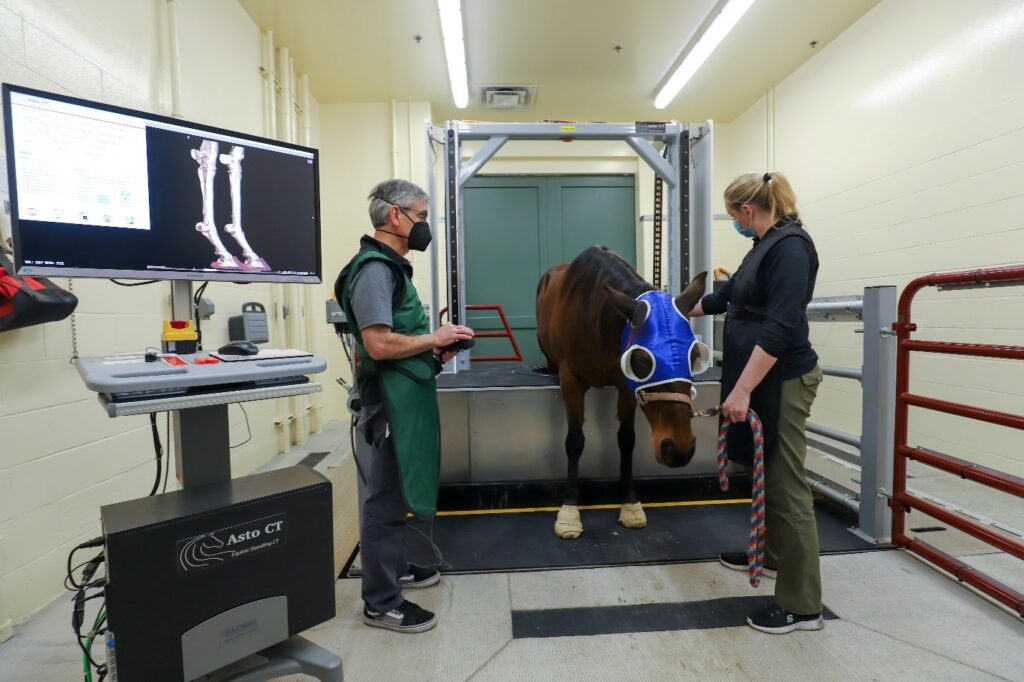
Traditionally horses need to be lying down when scanned and therefore, anesthetised for the process to take place. However Asto CT, a leader in equine diagnostic imaging technology, have developed the Equina Standing CT system. This minimises stress and risk to the horse, as no general anaesthetic is needed – an added bonus also that reduces cost considerably!
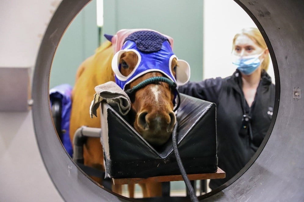
Standing CT allows the horse to remain upright and conscious with only light sedation during the imaging process. This not only reduces the risk associated with anesthesia but also, provides clearer and more precise images of the horse’s head, neck and limbs.
Designed to be non-invasive with the highest quality protection from the radiation leakage associated with radiographs, horses can be scanned standing naturally, which makes the whole procedure so much safer for all concerned.
What happens when a horse has a standing CT scan?
After a full work up to identify the problem area, the horse is lightly sedated then positioned within the scanner according to the body part to be examined – head, neck, fore or hind limbs. The whole process takes 10 -20 minutes from start to finish, with the actual scan lasting about 30 seconds while producing the most detailed images.
High quality images obviously, are essential for an accurate diagnosis – which include soft tissue injuries, subtle musculoskeletal issues, early signs of stress fractures or joint problems. Detailed 3D imaging from the get-go, enables vets to visualise the problem from all angles, so facilitating the best-informed decisions about a surgical procedure or, other treatment protocols.
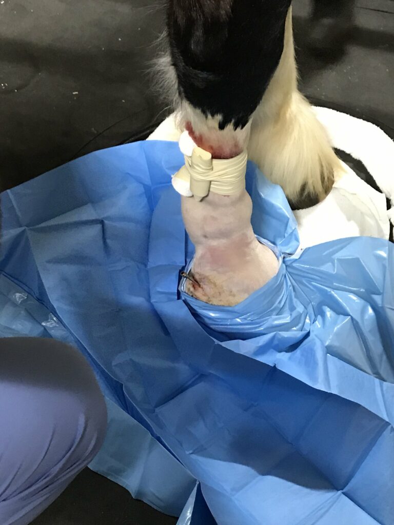
Case study
Shire gelding Roman, competes at the highest level on the show circuit. He was initially treated for an infection of the collateral cartilage to the outside of his foot. Although surgery went well, he returned to the clinic a couple of months later as healing was slower than expected, with a continually weeping wound.
CT investigation
It was recommended that Roman have a CT scan to investigate further. This showed a fistula – an abnormal tunnel-like tract – that connects between the skin and in this case, a bone sequestrum (a piece of dead bone that has separated from the healthy bone). When the body tries to expel the dead piece of bone surrounding it with infection, it creates a fistula. A bone sequestrum that is infected can delay the healing process and, will continue to drain and cause pain until it is removed.
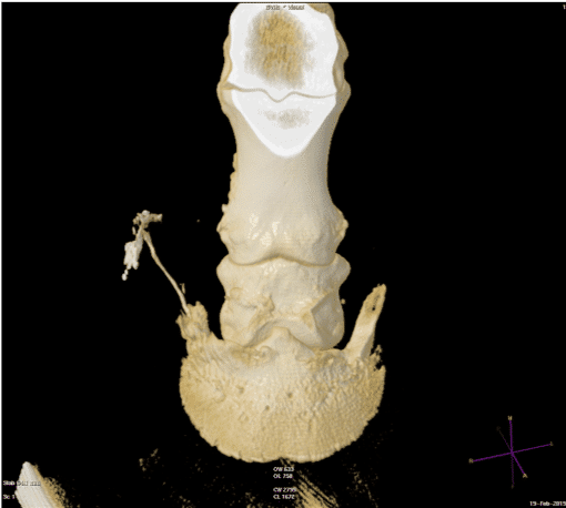
Roman’s CT images showed a draining tract from the coronary band that tracked all the way down to the coffin bone, to the suspected bone sequestrum.
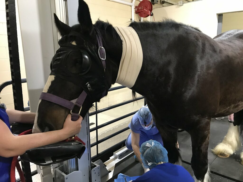
He was able to remain standing on top of the CT scanner while the surgical team were able to successfully debride the bone, using real time imaging. He is now doing much better at home.
Why is CT useful?
Interoperative imaging using the Equina standing CT scanner is particularly useful in cases where standing surgery is an option. CT imaging can be used to create 3D reconstructions of the affected area, which can be incredibly helpful in planning surgical procedures or interventions. In Roman’s case, these reconstructions allowed the team to assess the sequestrum’s relationship to other structures. They could then plan the best approach to minimise damage to healthy tissues and increase the chances of successful removal.
The Equina by Asto CT is set to be installed at Cotts Equine Hospital early next year with other Equine Hospitals set to install the scanner next year as well. Stay up to date with the latest news on Asto CT Equina installations by following their Facebook page: https://www.facebook.com/astoct/