Immune System, Veterinary
Why do we need to vaccinate horses?
Stuart Davies BVSc MRCVS, explains how and why vaccines for horses work
As horse owners we strive to maintain our horses in good health. One of the main ways we do this is by vaccinating against infectious pathogens – bacteria, virus or fungi that can cause disease. While there are many vaccines available for different diseases, who actually knows what, why or how the vaccine is producing immunity?
There are two pathways of the immune system that work together to protect the body from infection:
The innate immune system
The innate immune system is non-specific and comprises of anatomical (skin and mucous membranes), physiological, phagocytic (immune cell that can surround and kill microorganisms, ingest foreign material, and remove dead cells), and inflammatory barriers.
Pattern recognition receptors (PRRs) recognise common structures shared between molecules such as cell walls and viral RNA, Upon recognition, the innate immune system rapidly recruits immune cells to sites of infection or inflammation from the release of chemical signalling molecules called cytokines. This starts a cascade of chemical reactions both locally, in the tissue affected, and body wide to rid the pathogen from the body.
Macrophage cells can engulf the pathogen, break it down and also present the antigen, (a marker on the surface that enables the body to recognise it) to a T-cell.
The adaptive immune system
The adaptive immune system enables the body to recognise specific antigens and identify them as imposters, target their removal and, produce memory so that if reinfection occurs the body can recognise it swiftly. T cells are produced in bone marrow and circulate throughout the body in lymph fluid, (fluid that hydrates cells).
How is immunity acquired?
Immunity can be acquired from exposure to antigens in the environment. The foal is an example of this – it has come from a safe uterus into a world of antigens, which its body will have to recognise, determine if a risk, breakdown and get rid of, in addition to remembering it for the future. This is an example of active immunisation. This same process happens when we vaccinate an animal but, instead of using a pathogen able to produce infection it is often attenuated (weakened), inactivated or, only part of the pathogen.
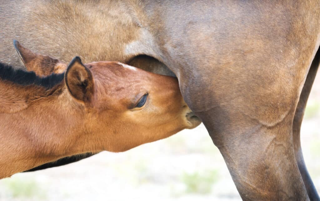
Foals have been shown to produce antibodies in utero. However, their immune system is not fully developed when born and in the first few weeks of life. A mare’s placenta actually prevents the transfer of the dam’s antibodies to the foal. Therefore, to help the foal deal with its environment, passive immunity is provided by the transfer of antibodies in colostrum.
Colostrum is produced by the mare, and only absorbed by the foal’s gut in the first 12-24 hours of life. The antibody that predominates in colostrum is IgG. Foals deficient in colostrum intake have been shown to be at a greater risk of infection. Therefore, it is commonplace for vets to perform an IgG test on a blood sample of a foal at roughly one day old. If there has been failure of passive transfer of maternally derived antibodies, either through inadequate colostrum of insufficient intake, then an infusion of hyperimmune plasma can be administered. The plasma given can be enriched with antibodies targeted against a specific pathogen such as Rhodococcus equi, a bacterial pathogen associated with pneumonia in foals.
Colostral antibodies, while helping the foal during immune system development, have been suggested to affect the success of vaccinations at a young age. Foals who received colostrum failed to produce antibodies following influenza vaccination until 6-8 months of age – when colostral antibodies declined. However, this mechanism is not fully understood. The same does not occur in adult horses who receive an influenza booster when they already have influenza antibodies from exposure or previous vaccination.
Vaccination timing
Vaccinations must be given within specific time periods between primary vaccination courses however, these time periods vary between the governing bodies of different countries. Interestingly, the window for 2nd vaccination for Equine Influenza Virus (EIV) can be 21-91 days after the first vaccination. In a study a greater number of individuals in the 2 and 3 month gap vaccine group, had insufficient antibody levels by the time of 2nd vaccination. However, in delaying the 2nd vaccination till the later period provided a more intense antibody peak, and, an increased time before antibody levels declined prior to booster vaccination a year later.
Vaccines for horses have been shown to reduce the likelihood of infection, spreading of the virus and, severity of clinical signs shown when infected. In outbreaks of EIV it becomes apparent that within vaccinated horses the older age group have a greater level of disease. This is due to the larger vaccination interval, often annual if not lapsed, in comparison to two-year olds who have received 4 vaccines within a two-year interval.
Vaccination site reactions
Vaccines are required to be updated as diseases mutate, so that protection applies to the strain currently circulating. However, some cross protection will be provided, but can’t be guaranteed from using vaccines containing historic strains.
In order to increase the recognition of the vaccine the attenuated, inactivated or, part vaccine, is mixed with an adjuvant, and it is the adjuvant that can cause an injection site reaction. This can present a pyrexia (fever), swelling and discomfort surrounding the injection site. Unfortunately, reducing the risk of injection site reaction, would affect bodily recognition and function of the vaccine. In summary, vaccinations are used to help protect the horse from infectious diseases.
References
- Dilai, M. et al. (2021) ‘An Evaluation of Three Different Primary Equine Influenza Vaccination Intervals in Foals’, Journal of equine veterinary science, 99, p. 103397. doi: 10.1016/j.jevs.2021.103397.
- Goehring, L.S., Wagner, B., Bigbie, R., Hussey, S.B., Rao, S., Morley, P.S. and Lunn, D.P. (2010) Control of EHV-1 viremia and nasal shedding by commercial vaccines. Vaccine 28, 5203-5211. Kahn, S. K. et al. (2019) ‘Transfusion With 2 L of Hyperimmune Plasma is Superior to Transfusion of 1 L or Less for Protecting Foals Against Subclinical Pneumonia Attributed to Rhodococcus equi’, Journal of Equine Veterinary Science, 79, pp. 54–58. doi: 10.1016/j.jevs.2019.05.015.
- Lai A.C., Chambers TM., Holland R.E. Jr, Morley PS., Haines D.M., Townsend H.C.G. & Barrendeguy M. (2001). – Diverged evolution of recent equine-2 influenza (H3N8) viruses in the Western Hemisphere. Arch. Virol, 146, 1063-1074. Perkins, G.A. and Wagner, B. (2015), The development of equine immunity. Equine Vet J, 47: 267-274. https://doi-org.liverpool.idm.oclc.org/10.1111/evj.12387
- Marshall, J.S., Warrington, R., Watson, W. et al. An introduction to immunology and immunopathology. Allergy Asthma Clin Immunol 14, 49 (2018). https://doi.org/10.1186/s13223-018-0278-1
- Van Oirschot, J.T., Bruin, G., de Boer-Luytze, E. and Smolders, G. (1991) Maternal antibodies against equine influenza virus in foals and their interference with vaccination. Zentralbl. Veterinarmed. B 38, 391-396.
- Watson, J. et al. (2011) ‘The 2007 outbreak of equine influenza in Australia: lessons learned for international trade in horses’, Revue scientifique et technique (International Office of Epizootics), 30(1), pp. 87–93. doi: 10.20506/rst.30.1.2021.