First Aid, Veterinary, Wound Care
What should I put on my horse’s wound?
Stuart Davies BVSc MRCVS, looks at topical wound dressings, why, when and what, should we use?
We have previously discussed the stages of equine wound healing to understand what steps need to be taken with wounds and, the possible reasons why a wound is slow or failing to heal. Here we will build on the brief mention of topical treatments and delve deeper into what and when to use common products.
Equine wounds often have a great degree of contamination when initially presented. Left in situ a small number of bacteria can colonise the wound, leading to ongoing inflammation and infection, that will spread from the wound site to affect the surrounding tissues. Ongoing infection is a major cause of non-healing wounds. If there is concern regarding any wound veterinary advice should be sought however, little harm can be done in conducting the first step of wound care whilst waiting for the vet to attend.
“Dilution is the solution to pollution”
Irrigation
Irrigation should be the first step when presented with a wound. ‘Dilution is the solution to pollution’ is a favourite saying; meaning that if large volumes of liquid are applied to the wound then there will likely be a reduction of bacterial load. Studies have shown that the ideal pressure of the irrigating solution is 7-15 psi, which is achieved by using a 35ml syringe and 19-gauge needle¹. However, a simple hose pipe can also be effective if the above equipment is not to hand. To add complexity, the solution used can have an influence on the wound.
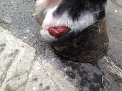
Figure 1. Overeach wound: A simple small and shallow overreach wound, with no damage to the coronet band, should initially be cleaned with dilute chlorhexidine or salt water prior to being lavaged with water.
Application of an antibacterial such as honey, or silver cream should then be applied under a light dressing. If you suspect injury to the coronary band, then management needs to be different and a vet should examine the wound.
Tap water
While being readily available, tap water is alkaline in Ph and can contain trace elements, both of which can be toxic to fibroblasts, the dividing cells involved in healing². Lactated ringers, or Hartmann’s solution; is the least contraindicated fluid for wounds². This solution is used as an intravenous fluid replacement and has the same osmolarity as cells in tissues. Sometimes clipping the haired wound edges and surrounding tissue is necessary for a thorough examination and cleaning. To prevent hairs from contaminating the wound a wound gel can be applied and removed once clipping has been completed.
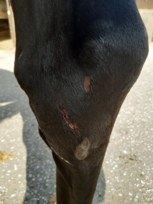
Figure 2: While these wounds may look minor in their size there are a lot of synovial structures (joints, tendon sheaths and bursa) around this location. Communication with synovial structures can have much worse consequences and require much more invasive treatment as opposed to aiming to healing the wound on its own.
Topical antiseptics
These can be used in conjunction with lavage fluids mentioned above and rid the wound of bacteria by both mechanical and chemical properties. Antiseptics need to be diluted to appropriate concentrations so as not to damage the fragile cells within the wound. The author utilises antiseptic solutions when first presented with a wound and with gross contamination. However, daily use on a healing wound should be avoided; they should NEVER be used without veterinary guidance.
Hydrogen peroxide’s usefulness is questionable. Upon contacting the wound it is broken down into oxygen and hydrogen, which inhibits bacterial killing. One possible use would be when Clostridium species of bacteria have colonised penetrative wounds as these rely on low oxygen environments to survive³. Hydrogen peroxide however, can cause skin burns and should not be used on an open wound and; certainly not without veterinary supervision.
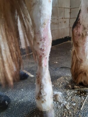
Figure 3: Wounds can commonly occur on white legs. UV light damages the skin’s immune barrier making self-trauma and other skin conditions commonplace. However, if your horse or pony is affected by multiple wounds then there could be a systemic problem. In this instance liver disease was diagnosed and skin changes were the only clinical sign.
Povidine-iodine works rapidly with a killing rate of 20-30 seconds for a lot of bacterial strains. However, it is inactivated by the presence of dirt and organic proteins, such as blood and serum, so can only be used once the wound has been irrigated⁴ .
Chlorhexidine is perhaps the most common antiseptic used by veterinarians for the initial cleansing of a wound. It has the benefit of working in the presence of organic material and dirt in comparison to iodine but, should not be used close to the eyes or when there is concern of communication with a joint due to the corneal damage and synovial inflammation it can cause ⁵.
Sodium hypochlorite, or bleach, has antibacterial properties hence its use in household cleaning. However, it is unstable and due to the array of less cytotoxic antiseptics available it should not be used for wound cleaning³.
Figure 3:This wound was managed open and healed well with copious twice daily application of silver ointment. Hypochlorous acid, an ingredient of many commercial skin cleansers, has low cytotoxic effects with good antibacterial activity. There is an over the counter and veterinary licensed formulation (Vetericyn®) which has also been recommended for the cleaning of chronic wounds⁶.
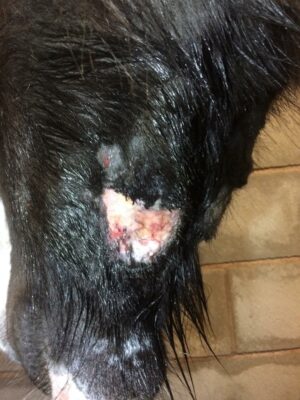
Figure 4: An abrasion like this head collar wound can heal quickly. A slight difficulty is closing wounds like this with sutures, because the tension present will likely cause the wound to reopen. A sticky dressing can be useful for keeping the wound covered.
Antimicrobial resistance
This is an emerging issue in both human and veterinary medical fields. With resistance emerging quicker than new antimicrobials being discovered, antimicrobials should be used judiciously to preserve the effectiveness. Antimicrobials can be applied topically to wounds as apposed to systemic (oral or injectable) administration. Silver sulfadiazine is a firm favourite for the author and colleagues as well as amongst human medical professionals in the treatment of burns⁷.
Cream preparations such as Flamazine® unfortunately do not adhere well to the wound and are inactivated over time therefore require daily application and the use of a light dressing. Honey is effective as an antimicrobial – medical grade manuka honey has been shown to help heal wounds on the distal limb of the foreleg (below the knee) up to 12 days faster compared to non-treated wounds⁸. It is useful during both the debridement and inflammatory phase of healing but, should not be used when healthy granulation tissue is present. The difficulties of applying this to the wound can be overcome with the use of available honey gels. Previously it has been suggested that the prolonged use of manuka honey leads to the production of exuberant granulation tissue, delaying wound healing. However, this has been disproven⁸.
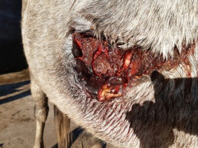
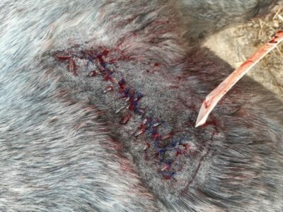
Figures 5 and 6 above: If you were presented with a wound like this it does unfortunately require veterinary assistance. The wound required debridement to remove old debris, thorough cleaning with chlorhexidine, lavage with hartmanns solution prior to being sutured closed. If left unsutured this wound would take a prolonged time to heal. Small pieces of tubing were used to reduce tension applied to the skin from the sutures, while a drain allowed any fluid to drain.
Exuberant granulation tissue
The most effective treatment for ‘proud flesh’ is by debridement and, best performed by a veterinarian. Despite the lack of nerve endings, sedation is advised for safety of everyone involved in-case of inadvertent contact to surrounding nerve filled tissue. Following sharp debridement corticosteroids can be topically applied to the wound underneath a dressing providing haemostasis to the richly vascular granulation tissue.
The exact mechanism of action is unknown but, as you have probably come to realise, like most topical products its use should be closely monitored, as it can inadvertently affect wound healing; specifically wound contraction, epithelialization (cell migration across the wound bed) and angiogenesis (blood vessel formation for the dividing cells of the wound bed).
Corticosteroids suppress the immune response so should not be used during the inflammatory phase of wound healing. Short term use is usually sufficient for preventing the recurrence of exuberant granulation tissue. The degree of systemic absorption is probably low, care must be taken in the treatment of large wounds in horses that may be steroid sensitive i.e., those with known endocrine issues, such as Equine Metabolic Syndrome, or Pituitary Pars Intermedia Dysfunction⁹.
Herbal products are widely available on the market for over-the-counter purchase by owners. A lot of their use is based on human evidence but is presumed to be similar in horses. Aloe vera is one such topical treatment, the main active ingredient being acemannan. It is reported to be soothing and stimulates the production of proinflammatory signalling chemicals within tissues inducing the proliferative phase of wound healing¹⁰.
Chamomile is another plant which contains a lot of therapeutic substances. However, the study compared its effect against wounds treated with corticosteroid, which as we know from above can have delaying effects on wound healing¹¹.
Tea tree oil has been shown to have both antimicrobial, antimicrobial properties. In addition, it contributes to fibroplasia so must be used conservatively and suitably diluted, during the inflammatory and proliferative phase of wound healing¹².
Arnica has been shown to have both anti-inflammatory and anti-purpuric (preventing blood vessel bursting and blood accumulation under the skin surface) effects. However, it is toxic when ingested so should only be used on intact skin making it more suited to contusions and of little use in equine wound healing¹³.
Dressings
Dressings can be useful when dealing with wounds of the distal limb (below the hock or knee). The primary dressing layer, which is in contact with the wound, can consist of foam or plastic film dressings depending on the degree of bleeding or exudation of the wound. An adherent surrounding rim can help keep the primary dressing in place. Various primary dressings are impregnated with antimicrobials or antibacterial products, such as manuka honey and silver. These aid the wound in its inflammatory debridement phase while maintaining an ongoing healthy granulation bed. A lack of movement and close observation of the horse’s comfort is imperative when a bandage is placed on a limb.
Pressure injuries can occur when bandages have been placed too tight or have slipped. The thickness of the bandage is determined by the likely movement of the skin in the location of the wound. In some cases, a Robert-Jones bandage is required to completely immobilise the area. As the wound heals, dressings can reduce in size and the possibility of a light dressing or elasticated alternative such as a presage can be used.
Hopefully the above information helps you assess or create a first aid kit to be used in the unfortunate event of an equine wound.
The above discussion is not exhaustive however in summary the author’s recommendation for your equine wound first aid kit:
- A foam dressing & cohesive bandage to achieve initial heamostasis if presented with an acutely bleeding wound.
- Sterile wound gel
- 1 litre of Hartmann’s solution
- 1 bottle of chlorhexidine, dilute 25ml in 1L of liquid to remove contamination in the initial wash
- Gauze swabs are preferential over cotton wool due to less particulates being shed into the wound
- Hydrogel
- Medical grade Manuka honey gel to be used in the initial debridement and inflammatory stages
- Silver sulfadiazine cream to be used when a healthy granulation bed is present
References
- A.J. Singer, J.E. Hollander, S. Subramanian, et al. (1994). ‘Pressure dynamics of various irrigation techniques commonly used in the emergency department’. Ann Emerg Med, 24, pp. 36-40
- E.A. Buffa, A.M. Lubbe, F.J. Verstraete, et al. (1997) ‘The effects of wound lavage solutions on canine fibroblasts: an in vitro study’ Vet Surg, 26 pp. 460
- Leise, B. S. (2018) ‘Topical Wound Medications’, Veterinary Clinics of North America: Equine Practice, 34(3), pp. 485–498
- D.E. Angel, P. Morey, J.G. Storer, et al. (2008) ‘The great debate over iodine in wound care continues: a review of the literature’ Wound Pract Res, 16, pp. 6-21
- D.G. Wilson, A.J. Cooley, P.S. MacWilliams, et al. (1994) ‘Effects of 0.05% chlorhexidine lavage on the tarsocrural joints of horses’ Vet Surg, 23, pp. 442-447
- C.L. Theoret, J. Schumacher . (2017) ‘Topical wound treatments and wound-care products’ (Eds.), Equine wound management (3rd edition), Wiley-Blackwell, Ames (IA), pp. 75-103
- Abanoglu H, Basaran O, Aydogan C, Azap OK, Karakayali F, Moray G. (2013) ‘Assessment of the effectiveness of silver-coated dressing, chlorhexidine acetate (0.5%), citric acid (3%), and silver sulfadiazine (1%) for topical antibacterial effects against the multi-drug resistant Pseudomonas aeruginosa infecting full-skin thickness burn wounds on rats’. Int Surg.98(4):416-23
- A.S. Bischofberger, C.M. Dart, N.R. Perkins, et al. (2013) ‘The effect of short- and long-term treatment with manuka honey on second intention healing of contaminated and noncontaminated wounds on the distal aspect of the forelimbs in horses’. Vet Surg, 42, pp. 154-160
- E. Farstvedt, T. Stashak, A. Othic. (2004) ‘Update on topical wound medications’. Clin Tech Equine Pract, 3, pp. 194-272
- S.F. Swaim, K.P. Riddell, J.A. Mcguire (1992) ‘Effects of topical medications on the healing of open pad wounds in dogs’ J Am Anim Hosp Assoc, 28, pp. 499-502
- Martins MD, Marques MM, Bussadori SK, et al. (2009) ‘Comparative analysis between Chamomilla recutita and corticosteroids on wound healing. An in vitro and in vivo study.’ Phytother Res; 23: 274
- Chin KB, Cordell B. (2013) ‘The effect of tea tree oil (Melaleuca alternifolia) on wound healing using a dressing model.’ J Altern Complement Med; 19: 942
- Reddy KK, Grossman L, Rogers GS. (2013) ‘Common complementary and alternative therapies with potential use in dermatologic surgery: risks and benefits.’ J Am Acad Dermatol; 68: e127