Anatomy, Diseases & Conditions, Featured, Skeletal
What is locking stifle in horses?
Ed Busuttil explains what happens when the horse’s stifle locks
The patellar lock mechanism provides an efficient way of keeping the limbs from flexing, meaning that horses can stand for an extended period of time without exerting too much energy. If the horse cannot replace their patella efficiently, resulting in it locking in place, the horse can appear lame and stiff. Before discussing the condition, it is essential to understand the anatomy and normal biomechanics of how the patellar lock mechanism works.

Normal anatomy – the basics
The stifle joint provides a key element within the stay apparatus of the horse. This is done by ‘hooking’ over the medial (inside) aspect of the femoral trochlea. The patella is lifted into position by a number of muscles which attach to the patella:
Directly – The quadriceps muscle, consisting of the vastus medialis, lateralis and intermedius and the rectus femoris¹.
Indirectly – sartorius, gracilis, tensor fasciae latae and biceps femoris².
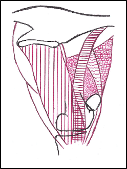
Figure 1: Schematic drawing showing the inside of the hindleg; showing the muscles which stabilise the stifle. From left to right, note the following muscles: gracilis (vertical lines), vastus medialis (circles), sartorius (horizontal lines), rectus femoris (wavy lines), tensor fascia latae (squares). Muscles generally provide 80% of the stabilisation of a joint.
Active stabilisation is necessary through the contraction of these muscles, especially the vastus medialis³.
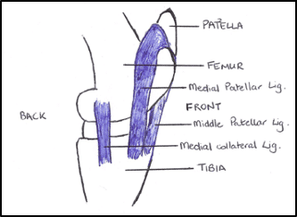
The patella is anchored to the tibia by three patellar ligaments – the lateral, middle and medial patellar ligaments. The medial patellar ligament is longer and weaker than the others⁴. Proximally, the vastus medialis inserts onto it⁴. As the patellar lock mechanism stabilises the whole limb distal to the hip, it is clear that the vastus medialis plays a key role in this stabilisation. If there is an anatomical variation to the medial femoral trochlea, or weakness to the muscles, which are involved in unlocking the stifle, the horse will be more likely to experience this disorder. (for a more detailed description of stifle anatomy see feature in Amazing Anatomy https://www.thehorsehub.co.uk/post/the-stifle)
Locking stifles
Horses which are predisposed to locking stifles include those which:
- Have an excessively straight hindlimb⁵
- Are not appropriately muscled or, have lost muscle tone following trauma or a period of box rest Hereditary factors may also be indicated, particularly in Shetland ponies⁶ Clinical signs depend on the severity and frequency of disease:
- Mild: Although lameness is not usually associated with this form⁷ there is a delayed patella release. If this becomes repetitive, it can cause some soreness. Partial locking may result in the hindlimb jerking up quickly, resembling stringhalt⁸.
- Moderate: In this form, the patella may remain locked for a few strides, an audible noise may be present when the patella is released.
- Severe: Horses with this form usually stand with the locked limb extended as the stifle and hock are prevented from flexing. It can last for a few minutes or be permanent, and happen on a frequent basis. Although some horses seem to accept it, others panic and try to release their leg⁹.
As the disorder can be intermittent, it may be difficult to diagnose. It can sometimes be provoked by trotting the horse up, backing up, circling in very tight circles towards the affected side, walking with a stop-start action or walking down a slope⁸.
The condition may be bilateral and, may be accompanied by swelling of the femoropatellar joint, particularly in chronic cases⁶, complicating the prognosis. This may also be altered depending on concurrent findings based on ultrasonographic and radiographic findings.
Treatment
Conservative
A conditioning programme to strengthen the muscles associated with the locking mechanism is generally initially indicated. Appropriate rehabilitation plans can be discussed with your veterinarian, chiropractor and RAMP registered practitioners, depending on the severity of the disease and following appropriate diagnosis, with increments in difficulty based on the progress of the horse.
Some exercises which could help to strengthen the stifle, through an improvement in conditioning, proprioception and coordination may include:
- Picking up the limb just above the fetlock and flexing it (like a flexion test), before slowly and safely pulling the limb backwards
- Long reining in straight lines
- Hydrotherapy
- Trot work over poles in an arc
- Walking over randomly placed poles
Increasing and improving the plane of nutrition, and ensuring that deworming and dental work is up to date is essential to ensure that any horse is receiving the right diet to condition appropriately.
More invasive techniques
These are performed by veterinary surgeons with the appropriate level of experience and expertise. They may be performed in the standing (sedated) horse or, under general anaesthetic, depending on the temperament of the horse and the veterinarian’s preference. The medial patellar ligaments are more easily palpated in the standing horse however, horse compliance is essential, both for the surgeon’s health and successful performance of the procedure. They should be performed when diagnosis is certain.
- Injection of counterirritants into and near the middle and medial patellar ligaments, potentially under ultrasound guidance. This causes severe ligament inflammation and fibroplasia¹⁰. A possible complication of this is accidental injection into the surrounding joints.
- Medial patellar ligament desmotomy⁶ involves, as the name implies, dissecting the medial patellar ligament. It may cause an instability within the femoropatellar joint and this can lead to subsequent damage to the cartilage and bone near where the ligament attaches to the patella. The horses need to be rested for 3-5 months to help the joint stabilise, and a persistent low-grade lameness, coupled with joint filling may be present.
- Medial patellar ligament splitting¹¹ involves inserting a surgical blade into the medial patellar ligament a number of times, to induce desmitis (swelling) so that the ligament is 2-3 times thicker. Horses begin exercise the following day, and although there are less side effects than the desmotomy procedure as the femoropatellar joint does not lose its stability, it is still very successful¹².
Thickening of the ligament is thought to stop the proximal part of the ligament from hooking over the femoral trochlea at the start of flexion, therefore preventing the disorder¹¹. Following the procedures, it is important to incorporate an appropriate rehabilitation plan.
Take home message
If you suspect that your horse or pony is suffering from a locking stifle, taking a good quality video of him or her when it happens could be a very useful aid for the veterinarian or qualified practitioner assessing your horse.
References
- Denoix, J. and Coudry, V., 2019. Ultrasonographic examination of the quadriceps femoris muscle: Normal images and findings. Equine Veterinary Education, 31(9), pp.478-482.
- Dyce, K., Sack, W. and Wensing, C., 1996. Textbook of Veterinary Anatomy. 2nd ed. Philadelphia: Saunders.
- Schuurman, S., Kersten, W. and Weijs, W., 2003. The equine hind limb is actively stabilized during standing. Journal of Anatomy, 202(4), pp.355-362.
- Sisson, S., 1975. Equine syndesmology. In: R. Getty, ed., The Anatomy of the Domestic Animals, 1st ed. Philadelphia: W.B. Saunders, pp.349-375. Rooney, J. and Robertson, J., 1996. Upward fixation of patella. In: J.
- Rooney and J. Robertson, ed., Equine Pathology, 1st ed. Ames: Iowa State University Press, p.203. Stashak, T., 2002. Upward patellar fixation. In: T.
- Stashak, ed., Adam’s Lameness in Horses, 5th ed. Philadelphia: Lippincott, Williams and Wilkins, pp.737-741.
- Dyson, S., 2002. Lameness associated with the stifle and pelvic regions. Proceedings of the American Association of Equine Practitioners, 48, pp.387-411.
- Tnibar, A., 2003. Treatment of upward fixation of the patella in the horse: an update. Equine Veterinary Education, 15(5), pp.236-242.
- Wyn-Jones, G., 1988. Upwards fixation of the patella. In: G. Wyn-Jones, ed., Equine Lameness, 1st ed. Oxford: Blackwell Scientific Publications, pp.170-174.
- van Hoogmoed, L., Agnew, D., Whitcomb, M., Hyde, D., MacDonald, M. and Snyder, J., 2002. Ultrasonographic and histologic evaluation of medial and middle patellar ligaments in exercised horses following injection with ethanolamine oleate and 2% iodine in almond oil. American Journal of Veterinary Research, 63(5), pp.738-743.
- Tnibar, M., 2002. Medial patellar ligament splitting for the treatment of upward fixation of the patella in 7 equids. Veterinary Surgery, 31(5), pp.462-467.
- Andersen, C. and Tnibar, A., 2016. Medial patellar ligament splitting in horses with upward fixation of the patella: A long-term follow-up. Equine Veterinary Journal, 48(3), pp.312-314.