Featured, First Aid, Veterinary, Wound Care
Abscesses, puncture wounds and tetanus in horses
Stuart Davies BVSc MRCVS, discusses the wounds that do need a vet visit …
Tetanus
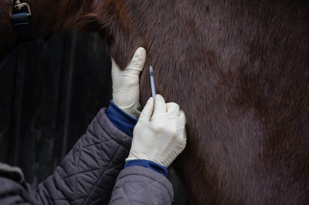
Primarily, one of the first questions in a vet’s mind will be the horse’s vaccination status. Horses in the UK are vaccinated against a multitude of diseases, including influenza and tetanus. The frequency of vaccinations may vary depending on the competition body you compete within. However, the primary course and vaccination frequency for tetanus protection remains the same. Having seen several cases of fatal tetanus in unvaccinated individuals so far in my career, I would urge everybody to vaccinate against tetanus to prevent any equid suffering from this agonising, paralysing condition.
What causes tetanus?
Clostridium tetani is a bacterium that produces spores, which lay dormant in the environment for some time. The bacterium/ spores gain entry to the body usually via soil contamination of a wound (accidental or surgical) following which it produces a neurotoxin. This neurotoxin attacks the nerves within the body. The time between infection and appearance of clinical signs is usually between 7 and 21 days. However, once clinical signs manifest the disease is often fatal in 5-10 days¹
Symptoms of tetanus in horses:
- Excessive sensitisation to touch, light and sound
- Limb & neck stiffnes Involuntary muscle contractions
- Inability to move jaws
- Tail held up and out
- 3rd eyelid positioned over eyeball rather than within inner corner
The disease progresses to the horse being unable to stand or breath, profusely sweating and inability to swallow¹. It is vital for owners to be aware of the clinical signs of the disease as diagnosis is often based on these, in addition to a history of a wound/ surgery, in an unvaccinated individual.
Rapid and aggressive treatment is vital with chances of survival reported to be approximately 50%². This involves hospitalisation to provide antibiotics, muscle relaxants, sedation and tetanus antitoxin. Any wounds that are present are cleaned with antiseptics to remove any contamination as best as possible. Due to sweating and inability to eat an electrolyte imbalance needs to be corrected using fluid therapy and the horse maintained in a comfortable and quiet environment³.
How do we prevent this devastating disease?
VACCINATE! Many people disagree with the frequency of booster vaccinations amongst horses, with comparisons made to the long duration of immunity amongst people. One study showed that duration of immunity after the first 3 doses lasted at least for 3 years⁴.
Additional protection to mares about to foal can be given by a booster in the last month of pregnancy. This has the added benefit of providing antibodies via the colostrum to the newborn foal, to protect them up until their first vaccination at 4-6 months. If foals are born to an unvaccinated mare, then vaccines should be performed at an earlier age.
Puncture wounds
A puncture wound poses a particular risk of concern due to the inability to estimate the extent of internal damage from mere external examination. Previous discussion focused on the delayed healing of a wound and in penetrative wounds, this can be a particular nuisance. The skin wound itself is often narrow and can heal prior to any material at the depths of the wound being extruded. In addition, bacteria such as Clostridium tetani, as discussed above, can be taken into the wound causing infection and associated complications.
The small appearance of the skin wound associated with a puncture wound can make an owner aloof to the seriousness of the injury. Wounds can extend much much deeper through subcutaneous tissues and addressing this quickly is imperitive. Infection within a joint can be fatal and those with better luck, or with infection in the surrounding structures, can develop early onset severe arthritis.
To determine the depth of these injuries imaging needs to be performed. Radiography, ultrasonography and magnetic resonance imaging (MRI) all can be used to identify damaged structures or remnants of the foreign object. If the object is no longer in place, a sterile probe can be inserted into the wound or, a joint can be distended with saline to determine if the wound communicates with the joint. The small skin wound often prevents thorough debridement and retrieval from this aspect so often surgery is required.
“if your horse has had a penetrative wound and becomes uncomfortable on that limb veterinary examination is a necessity!”
Infection of synovial structures such as joints, tendon sheaths and bursa are associated with a severe non-weight bearing lameness. Therefore, if your horse has a history of a penetrative wound and becomes uncomfortable on that limb veterinary examination is a necessity! If material remains within the depths of a wound, or a synovial structure/ tendon/ bone is contaminated, surgery is required to remove this potential for ongoing infection. Following which systemic (injectable/ oral) or regional antibiotic perfusion is required. For the lower limb, a tourniquet can be applied to occlude blood return and delivery to the extremities. Antibiotic can then be instilled into the vessels in the area prior to the tourniquet being removed.
So… you’ve found your horse with a puncture wound, what can be done while the vet is on the way?
Firstly, if the object is still in place attempt to secure it so it doesn’t break, bend or penetrate further. If the object has been removed then clip the area so that no obstruction to drainage is present, clean the surface of the wound with dilute hibiscrub and apply some sort of sterile dressing to prevent further contamination.
Hoof abscess
A differential, and much more common, diagnosis for an acute severely lame horse is a hoof abscess. This is usually due to a defect within the wall-sole junction or white line that allows bacteria/ debris to penetrate the hoof complex. Inflammation is initiated and as white blood cells and fluid accumulates, pain experienced by the horse rapidly increases from pressure over the underlying solar layers.
Once hoof testers have been used to identify an area of discomfort the white line can be parred away to open the defect, hopefully, to relieve greyish pus. It is likely that the pus will spread under the adjacent sole as it passes through the route of least resistance. However, care should be taken to not remove sole, creating a portal of entry for further bacteria and causing damage to the deeper corium, the vessel network supplying blood to the hoof structure.
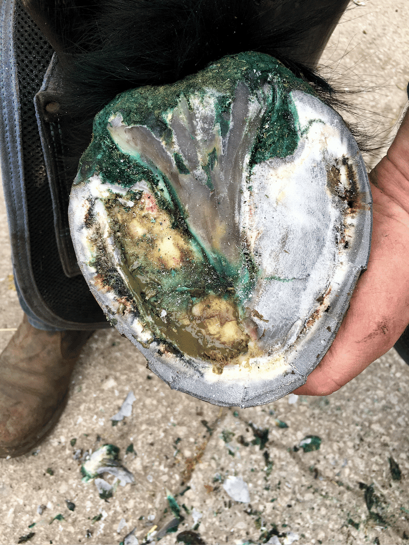
Hoof abscesses while incredibly common can come with associated complications. Deeper structures such as the pedal bone can become infected, termed osteomyelitis, requiring a greater intervention than merely establishing drainage. If drainage isn’t quickly established pus tracks up and exudes at the coronet band. While this can provide some improvement in comfort for the horse with the release of pressure, it can lead to a longer recovery time, recurrence of abscesses and hoof wall cracks⁵.
A medicated poultice can be applied whether/ not drainage has been established. Epsom salts can be used on poultice material to enhance antibacterial properties and osmotic draw. Once pus has ceased, approximately 3 days, the defect must be protected from repeated infection. This can be done using a dry poultice. For more information on how and when to poultice follow the link How and when should I use a poultice.
Recurrence of abscesses could be due to a multitude of reasons and require a conversation with your vet:
- Poor hoof horn quality or care: feeding of 20milligrams of biotin daily improved horn quality after 9 months⁶
- Mixed weather conditions affecting hoof softness Inadequate drainage established initially
- Subclinical laminitis
- Pedal bone osteomyelitis
- Keratoma
References
- Mackay R.J. 2014. Tetanus, p.368-372. In: Sellon D.C. & Long M.T. (Eds), Equine Infectious Diseases. 2nd ed. Saunders Elsevier, St Louis, Missouri.
- Reichmann P., Lisboa J.A.N. & Araújo R.G. 2008. Tetanus in equids: a review of 76 cases. J. Equine Vet. Sci. 28(9):518-523. http://dx.doi.org/10.1016/j.jevs.2008.07.019.
- Kay G. & Knottenbelt D.C. 2007. Tetanus in equids: a report of 56 cases. Equine Vet. Educ. 19(2):107-112. http://dx.doi.org/10.2746/095777307X181320.
- Kendall, A., Anagrius, K., Gånheim, A., Rosanowski, S.M. and Bergström, K. (2016), Duration of tetanus immunoglobulin G titres following basic immunisation of horses. Equine Vet J, 48: 710-713. https://doi-org.liverpool.idm.oclc.org/10.1111/evj.12502
- Redding, W. R. and O’Grady, S. E. (2012) ‘Septic Diseases Associated with the Hoof Complex: Abscesses, Punctures Wounds, and Infection of the Lateral Cartilage’, Veterinary Clinics of North America: Equine Practice, 28(2), pp. 423–440.
- Josseck, H., Zenker, W. and Geyer, H. (1995) ‘Hoof horn abnormalities in Lipizzaner horses and the effect of dietary biotin on macroscopic aspects of hoof horn quality’, Equine veterinary journal, 27(3), pp. 175–182.