Colic, Veterinary
What are the types of colic?
Dr Emma Shipman, BVetMed DipACVIM Cert VA MRCVS, University of Nottingham Vet School & Oakham Veterinary Hospital explains all
Colic is the term used to describe pain associated with the abdomen (belly). This may involve the urinary tract (kidneys and bladder) and the reproductive organs but, is generally associated with pain caused by abnormalities in the gastrointestinal tract (GI tract or gut).
The GI tract briefly is consisted of the oesophagus, stomach, small intestine, large intestine and rectum. In horses the large intestine is specially adapted for bacterial fermentation of grass and, consists of a tube folded into a double V shape, with a blind ending sac (the caecum), at the junction between the small and large intestine. As an estimate of large intestine size, the capacity of the stomach in an adult 500kg horse is approximately 12-15 litres. The combined capacity of the caecum and large colon in the same horse would be approximately 140-150 litres.
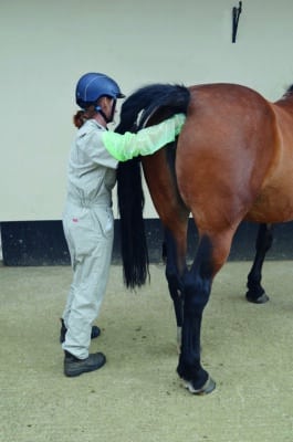
Figure 1. Rectal exam by a veterinary surgeon. (Image courtesy University of Nottingham)
The intestinal tract is held in place within the abdomen by a thin layer of tissue called the mesentery. It is within this mesentery that the nerves and blood vessels travel to different parts of the gut. In some regions the mesentery is short allowing little movement of the gut. In some regions the mesentery is much longer allowing much more free movement of the GI tract. This is the case for much of the small intestine, which can move relatively freely with the abdomen along much of its length. In some regions, for example the pelvic flexure (one of the U bends in the folded large colon), the gut in not attached by mesentery at all, leaving it able to move within the abdomen.The GI problems that result in colic signs can generally be divided into 4 categories: impactions, displacements, torsions or twists, and motility disorders (changes in the gut movement).
Impactions
Impactions occur when food (ingesta) moving along the GI tract becomes dry and solid, and unable to pass along the intestine. Further ingesta meeting the obstruction cannot continue to pass and, the stretching of the gut associated with this impaction causes pain and colic signs. Impactions can occur in a number of places along the GI tract but in the UK, the most common site is the pelvic flexure of the large colon. This is readily palpable on rectal examination as a large doughy structure and is one of many reasons why your veterinary surgeon will commonly carry out an examination per rectum with a horse showing colic signs. Other sites that can develop impactions include other U bends within the large colon (sternal and diaphragmatic flexures) the caecum, the small colon (the last part of the large intestine before the rectum) and the ileum (the last part of the small intestine).
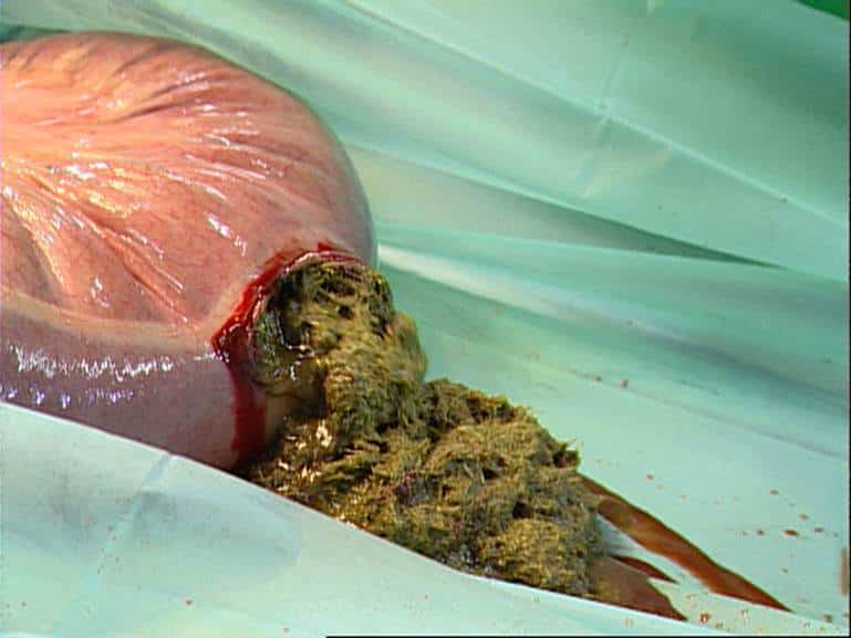
Figure 2
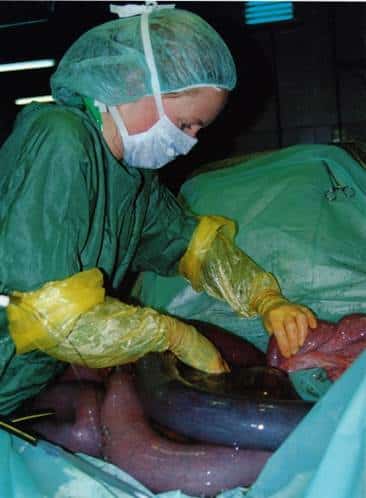
Figure 3
Images above: Figure 2. Incision into the pelvic flexure (large colon) to remove contents during surgery for a large intestinal displacement and impaction. (Image courtesy University of Nottingham). Figure 3. Surgery for a small intestinal strangulation. (Image courtesy Professor Sarah Freeman, University of Nottingham).
Spasmodic colic
This is a wide-ranging term for horses presenting with colic where other abnormalities cannot be found, and, which generally have increased gut movement (and therefore gut noise if you listen over the belly). The colic signs are associated with increased gut spasm due to the increase in motility (a horse equivalent to gut cramps that we may experience after a very spicy curry for example). Rectal examination is within normal limits in these cases and, these horses often respond very favourably to drugs that decrease gut motility (see treatment of colic).
Displacements
Displacements involve movement of parts of the GI tract into positions where they are not usually found. The large colon is the structure most commonly displaced. If gas builds up from the bacterial fermentation within the colon that cannot escape, this tends to accumulate causing gut stretching and pain, and also wedging the gut more firmly in its displaced location. This gut stretch and gas accumulation are, again, often palpable on rectal examination.
Gut torsion or twisting
This type of colic is often associated with the most severe signs of pain (sometimes of uncontrollable severity) and, with the urgent requirement for timely examination of a horse with colic as soon as signs of abdominal pain are present. With gut torsion the gut twists about its long axis and, this results in blockage of arterial and venous blood supply to the affected region of gut. Without a blood supply to deliver oxygen the cells of the intestine start to die.
Not only is this process painful but, the adhesions between the dying gut cells break down and become leaky. This allows the bacteria normally within the gut and their associated toxins to leak into the blood stream, leading to development of severe generalised inflammation (sepsis or blood poisoning) and subsequent shock. Colic caused by gut torsion requires surgical treatment.
For further information on a wide range of issues related to colic visit bhs.org.uk/colic
Oakham Veterinary Hospital is a BHSVet REACT Colic Champion